Cardiac Magnetic Resonance Imaging

2023| Myocardial Inflammation at FDG PET/MRI and Clinical Outcomes in Symptomatic and Asymptomatic Participants after COVID-19 Vaccination

150 150 fraser.amos@uhn.ca

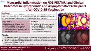
Authors: Constantin Arndt Marschner, Paaladinesh Thavendiranathan, Dakota Gustafson, Kathryn L. Howe, Jason E. Fish, Robert M. Iwanochko, Rachel M. Wald, Husam Abdel-Qadir, Slava Epelman, Angela M. Cheung, Rachel Hong, Kate Hanneman

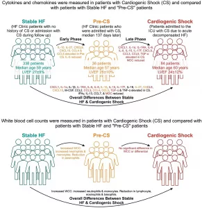
Short Description: As of August 2022, more than 5.3 billion people worldwide have received at least one dose of a COVID-19 vaccine (1). Several adverse events have been reported, including myocarditis and pericarditis, following the administration of mRNA-based COVID-19 vaccines (2,3). The overall incidence of myopericarditis following COVID-19 vaccines is low, estimated at 18 per 1 million vaccine doses, with the highest risk in adolescent and young adult males (4). However, many more patients experience cardiac symptoms after vaccination, including shortness of breath, palpitations, and chest pain, yet do not meet diagnostic criteria for acute myopericarditis (5). The cause of these symptoms and the natural history of these patients remains unknown.

Cardiac MRI plays an important role in the assessment of myocardial tissue changes (6). Cardiac fluorine 18 (18F) fluorodeoxyglucose (FDG) PET provides complementary physiologic information, allowing for the assessment of changes in myocardial metabolism (7,8). Several recent case series have reported cardiac MRI findings in patients with myocarditis following COVID-19 vaccination (9,10). However, there are limited data on patients who present with new-onset cardiac symptoms following COVID-19 vaccination but do not meet diagnostic criteria for myopericarditis (5). There are also limited data evaluating potential subclinical myocardial tissue changes in asymptomatic individuals after vaccination.

The purpose of this study was to identify cardiac sequelae of COVID-19 vaccination and relate patient-reported cardiac symptoms to myocardial tissue changes identified with cardiac FDG PET/MRI, circulating biomarkers of cardiac injury and systemic inflammation, health-related quality of life, and adverse cardiac events at 2-month follow-up. Our goal was to inform guidelines for cardiac investigations after COVID-19 vaccination and the potential need for longer-term follow-up.

Interest: Myocardial Inflammation, COVID-19, Vaccination, Molecular Imaging, MR Imaging

Click Here to Read the Full Publication

2025 | Heart Failure Decompensation with Cardiogenic Shock Exhibits Distinct Sequential Inflammatory Profiles

150 150 fraser.amos@uhn.ca

Authors: Darshan H. Brahmbhatt, Fernando Luis Scolari, Nicole L. Fung, Madison Otsuki, Patrick R. Lawler, Heather J. Ross, Uros Kuzmanov, Anthony O. Gramolini, Adriana C. Luk, Filio Billia

Short Description: Cardiogenic shock (CS) is the most severe form of acute heart failure (HF) characterized by severely reduced cardiac output and inadequate end-organ perfusion leading to tissue hypoxia. Patients with CS suffer significant morbidity and mortality1 with in-hospital mortality reaching up to 50%.2 The pathophysiological underpinnings of CS remain to be fully elucidated, but inflammation is thought to play a key role.3 One of the several potential mechanisms for worsening of the shock state has been proposed to occur through the release of inducible nitric oxide and its mediators, resulting in inappropriate vasodilatation.4 Additionally, the development of systemic inflammatory response syndrome (SIRS) is seen in up to a third of patients at presentation and portends a more severe clinical syndrome associated with worse outcomes.5

The inflammatory profile associated with CS is now being studied in greater detail. However, the majority of studies have focused on CS following acute myocardial infarction (AMI-CS). In AMI-CS, an acute ischaemic insult leads to an immediate drop in cardiac output, which may then be compounded by the ischaemia–reperfusion injury associated with acute revascularization.6 In contrast to this, CS caused by acute decompensated HF (ADHF-CS) often has a heterogeneous trajectory, even before presentation. This realization is important in that these patients may have a different clinical profile and outcomes after discharge.7 The HF state has its own heightened inflammatory state, which is also known to affect outcomes.8 Thus far, the inflammatory profile of patients with ADHF-CS has not been fully characterized nor has the transition from a clinically stable, outpatient-managed HF state to ADHF-CS, and it is unclear if there are changes in the inflammatory profile that occur during this clinical deterioration.

This retrospective, cross-sectional study was designed to investigate the profile and prognostic significance of circulating cytokines and cellular inflammatory profiles in patients with ADHF-CS admitted to a cardiac intensive care unit (CICU) and to define the association between circulating cytokines in outpatients with HF and those admitted with CS.

Interest: cardiogenic shock, heart failure, cytokines, mechanical circulatory support, biomarkers, chemokines

Click Here to Read the Full Publication

2025 | Pretreatment Circulating Vascular Biomarkers Predict Cancer Therapy-Related Cardiac Dysfunction During HER2+ Breast Cancer Treatment

150 150 fraser.amos@uhn.ca

Authors: Dakota Gustafson, Priya Mistry, Crizza Ching, Inbar Nardi-Agmon, Christopher Yu, Chun-Po Steve Fan, Christian Houbois, Eitan Amir, Thomas Marwick, Husam Abdel-Qadir, Chris McIntosh, Paaladinesh Thavendiranathan, Jason E Fish

Short Description:Blood biomarkers to predict cancer therapy-related cardiac dysfunction (CTRCD) risk remain limited. The aim of this study was to identify circulating biomarkers associated with CTRCD risk in HER2+ breast cancer patients. Pretreatment endothelial-centric and inflammatory biomarkers outperformed both clinical and CMR measures in predicting CTRCD during chemotherapy.

Interest: angiopoeitin-2, biomarkers; breast cancer; cancer therapy–related cardiac dysfunction; cardio-oncology; endothelium; inflammation; machine learning; microRNA; myeloperoxidase; vascular

Click Here to Read the Full Publication

2023 | Large scale genome-wide association analyses identify novel genetic loci and mechanisms in hypertrophic cardiomyopathy

150 150 sabrina.agostini@uhn.ca

Authors: Rafik Tadros, Sean L. Zheng,  Christopher Grace, Paloma Jordà, Catherine Francis, Sean J. Jurgens, Kate L. Thomson Andrew R. Harper, Elizabeth Ormondroyd, Dominique M. West, Xiao Xu, Pantazis Theotokis, Rachel J. Buchan, Kathryn A. McGurk, Francesco Mazzarotto, Beatrice Boschi, Elisabetta Pelo, Michael Lee, Michela Noseda, Amanda Varnava, Alexa Mc Vermeer, Roddy Walsh, Ahmad S. Amin, Marjon A van Slegtenhorst, Nicole Roslin, Lisa J. Strug, Erika Salvi, Chiara Lanzani, Antonio de Marvao, Hypergenes InterOmics Collaborators, Jason D. Roberts, Maxime Tremblay-Gravel, Genevieve Giraldeau, Julia Cadrin-Tourigny, Philippe L’Allier, Patrick Garceau, Mario Talajic, Yigal Pinto, Harry Rakowski, Antonis Pantazis, John Baksi, Brian P. Halliday, Sanjay K. Prasad, Paul Jr Barton, Declan P. O’Regan, Stuart A. Cook, Rudolf A. de Boer, Imke Christiaans, Michelle Michels, Christopher Kramer, Carolyn Y. Ho, Stefan Neubauer, HCMR Investigators, Paul M. Matthews, Arthur A. Wilde, Jean-Claude Tardif, Iacopo Olivotto, Arnon Adler, Anuj Goel, James S. Ware, Connie R. Bezzina, Hugh Watkins

Short Description: Hypertrophic cardiomyopathy (HCM) is an important cause of morbidity and mortality with both monogenic and polygenic components. We here report results from the largest HCM genome-wide association study (GWAS) and multi-trait analysis (MTAG) including 5,900 HCM cases, 68,359 controls, and 36,083 UK Biobank (UKB) participants with cardiac magnetic resonance (CMR) imaging. We identified a total of 70 loci (50 novel) associated with HCM, and 62 loci (32 novel) as sociated with relevant left ventricular (LV) structural or functional traits. Amongst the common variant HCM loci, we identify a novel HCM disease gene, SVIL, which encodes the actin-binding protein supervillin, showing that rare truncating SVIL variants cause HCM. Mendelian randomization analyses support a causal role of increased LV contractility in both obstructive and non-obstructive forms of HCM, suggesting common disease mechanisms and anticipating shared response to therapy. Taken together, the findings significantly increase our understanding of the genetic basis and molecular mechanisms of HCM, with potential implications for disease management.

Interest: Cardiac Magnetic Resonance Imaging, Cardiac Imaging, Genetics, Genetic Testing, Hypertrophic Cardiomyopathy

Click Here to Read the Full Publication

2022 | Temporal Changes in Cardiac Morphology and Its Relationship with Clinical Characteristics and Outcomes in Patients with Hypertrophic Cardiomyopathy

150 150 sabrina.agostini@uhn.ca

Authors: Manhal Habib, Arnon Adler, Sara Hoss, Kate Hanneman, Olga Katz, Hadeel Halloun Habib, Kimia Fardfini, Harry Rakowski, Raymond H. Chan

Short Description:  In this study, we aimed to assess a large cohort of nonapical hypertrophic cardiomyopathy (HC) patients who have undergone 2 serial cardiac magnetic resonance studies to examine morphological dynamics and their correlation to patient characteristics and clinical outcomes. A total of 214 patients with nonapical HC were enrolled in this study, with 2 sequential cardiac magnetic resonance studies separated by a mean interval of 4.8 ± 2.1 years.

Interest: Hypertrophic Cardiomyopathy

Click Here to Read the Full Publication

2022 | Importance of newer cardiac magnetic resonance–based risk markers for sudden death prevention in hypertrophic cardiomyopathy: An international multicenter study

150 150 sabrina.agostini@uhn.ca


Authors: Ethan J. Rowin, Martin S. Maron, Arnon Adler, Alfred J. Albano, Armanda M. Varnava, Danna Spears, Dana Marsy, Stephen B. Heitner, Emilie Cohen, Kevin M.W. Leong, Stephen L. Winters, Matthew W. Martinez, Benjamin C. Koethe, Harry Rakowski, Barry J. Maron

Short Description: The sudden death (SD) risk stratification algorithm in hypertrophic cardiomyopathy (HCM) has evolved, underscored recently by novel cardiac magnetic resonance (CMR)–based risk markers (left ventricular apical aneurysm, extensive late gadolinium enhancement, and end-stage disease with systolic dysfunction) incorporated into the 2020 American Heart Association (AHA)/American College of Cardiology (ACC) HCM guidelines. The purpose of this study was to assess the specific impact of newer, predominantly CMR-based risk markers in a large multicenter HCM population that underwent primary prevention implantable cardioverter-defibrillator (ICD) implants.

Interest: Cardiac Imaging, Cardiac Magnetic Resonance Imaging, Hypertrophic Cardiomyopathy, Medical Imaging, Risk Stratification, Sudden Death

Click Here to Read the Full Publication

2021 | Left Ventricular Apical Aneurysms in Hypertrophic Cardiomyopathy: Equivalent Detection by Magnetic Resonance Imaging and Contrast Echocardiography

150 150 sabrina.agostini@uhn.ca

Authors: Deacon Z.J. Lee, Raymond H. Chan, Mahdi Montazeri, Sara Hoss, Arnon Adler, Elsie T. Nguyen, Harry Rakowski 

Short Description: Left ventricular (LV) apical aneurysm is a unique morphological entity and novel adverse risk marker existing within the broad phenotypic spectrum of hypertrophic cardiomyopathy (HCM). Its true prevalence in the HCM population is likely underestimated because of inherent limitations of conventional noncontrast echocardiography. The authors hypothesized that contrast echocardiography is a reliable imaging technique compared with cardiovascular magnetic resonance (CMR) for the detection of apical aneurysms. The aim of this study was to assess the effectiveness of contrast echocardiography in the detection of LV apical aneurysms in patients with HCM in comparison with the gold standard, CMR.

Interest: Cardiac Imaging, Cardiac Magnetic Resonance Imaging, Hypertrophic Cardiomyopathy, Left Ventricular Apical Aneurysm, Medical Imaging

Click Here to Read the Full Publication

2021 | Progression of Myocardial Fibrosis in Hypertrophic Cardiomyopathy: A Cardiac Magnetic Resonance Study

150 150 sabrina.agostini@uhn.ca

Authors: Manhal Habib, Arnon Adler, Kimia Fardfini, Sara Hoss, Kate Hanneman, Ethan J. Rowin, Martin S. Maron, Barry J. Maron, Harry Rakowski, Raymond Chan

Short Description: Myocardial fibrosis, as quantified by late gadolinium enhancement (LGE) in cardiac magnetic resonance (CMR), provides valuable prognostic information in patients with HCM. This study examined fibrosis progression in hypertrophic cardiomyopathy (HCM) patients, as well as its relationship to patient characteristics, clinical outcomes, and its effect on clinical decision making.

Interest: Cardiac Imaging, Cardiac Magnetic Resonance Imaging, Hypertrophic Cardiomyopathy, Medical Imaging

Click Here to Read the Full Publication

Privacy Preferences

When you visit our website, it may store information through your browser from specific services, usually in the form of cookies. Here you can change your Privacy preferences. It is worth noting that blocking some types of cookies may impact your experience on our website and the services we are able to offer.

Click to enable/disable Google Analytics tracking code.
Click to enable/disable Google Fonts.
Click to enable/disable Google Maps.
Click to enable/disable video embeds.
Our website uses cookies, mainly from 3rd party services. Define your Privacy Preferences and/or agree to our use of cookies.